|
|
||||||||||||||||||
Modality-Classification of Microscopy Images Using Shallow Variants of Deep Networks
Authors: Trelles Trabucco, J, Li, P,, Arighi, C., Shatkay, H., Marai, G.E.
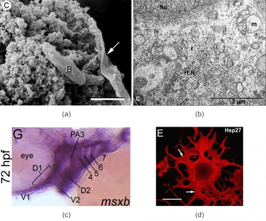
Publication: 2020 IEEE International Conference on Bioinformatics and Biomedicine (BIBM) Microscopy images are pervasive in biomedical research publications, where images obtained through various microscopy modalities (light, fluorescence, scanning, transmission) are often used to describe and summarize experiments and contributions. Hence, there is growing interest in automatically identifying these microscopy images’ modality and utilizing this knowledge in automated search tools. However, identifying microscopy images poses challenges due to a lack of extensive collections of labeled images. We describe and evaluate two alternative approaches to microscopy image classification. In the first approach, we progressively fine-tuned layers of ResNet models. The second approach uses shallow variants of ResNet networks, where we leverage the outputs from previous convolutional blocks. We compare these results against a Support Vector Machine (SVM)-based baseline. Our results show that fine-tuning specific layers yields better results than fine-tuning the whole model. Furthermore, shallower variants produce competitive results when compared to the entire fine-tuned model. Index Terms: microscopy, image classification, deep learning Date: December 16, 2020 - December 19, 2020 Document: View PDF |